Introduction
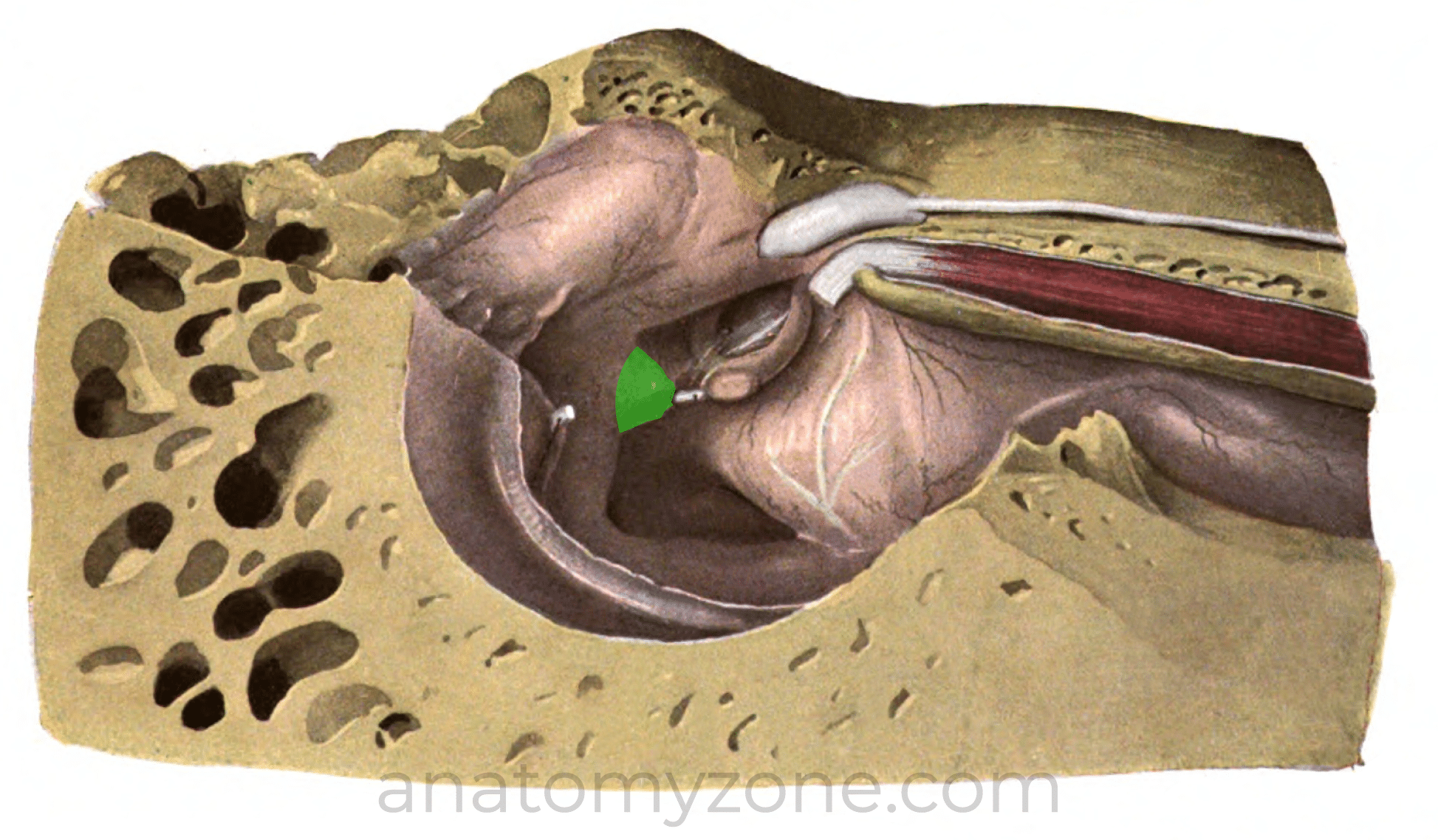
There are 3 ossicles which bridge the middle ear cleft, attaching laterally to the tympanic membrane and medially to the oval window of the inner ear, the malleus, the incus and the stapes.
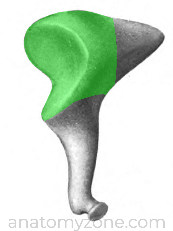
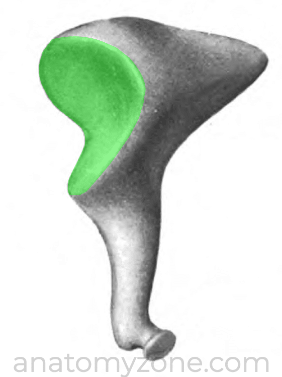
Malleus
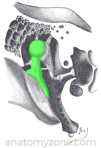
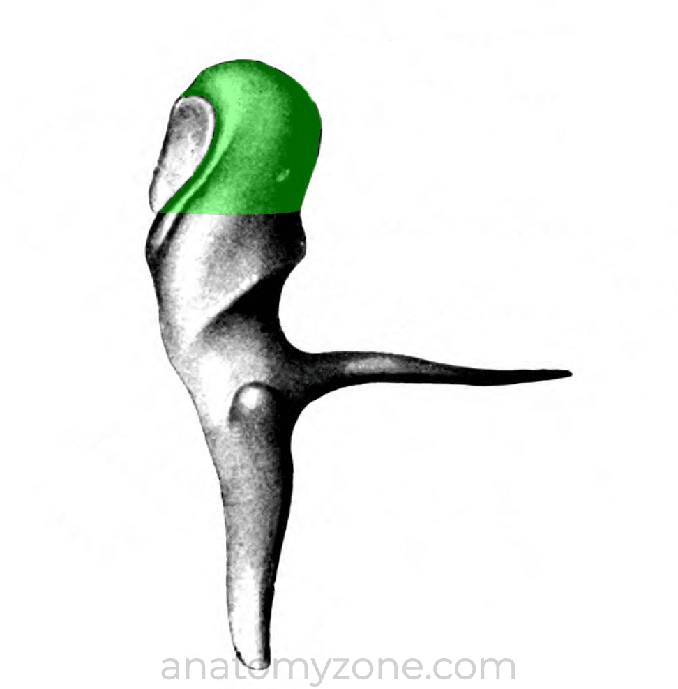
The malleus derives its name from its hammer shape and is the first bone in the ossicular chain, which attaches to the inner surface of the tympanic membrane.
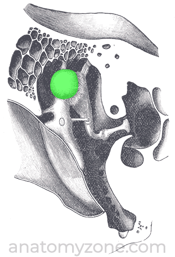
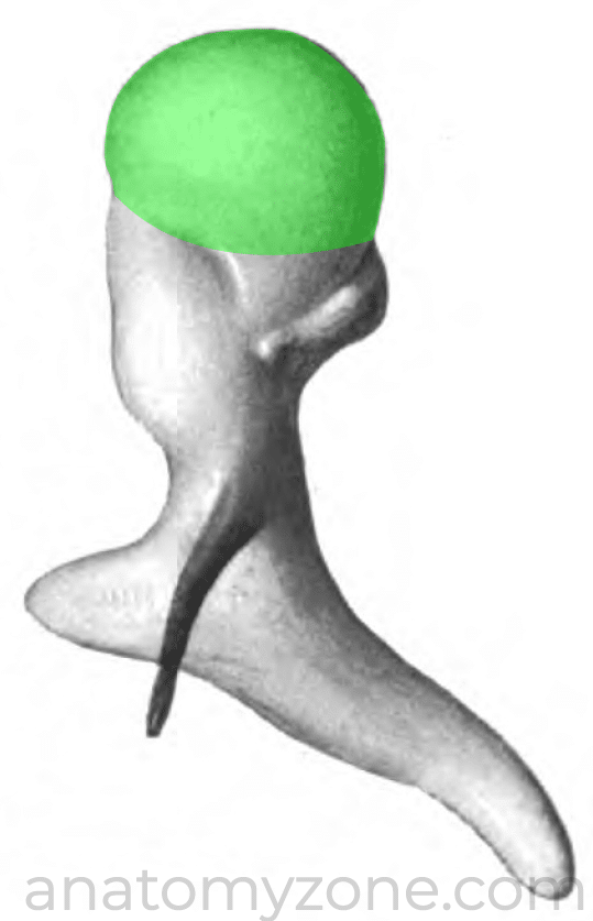
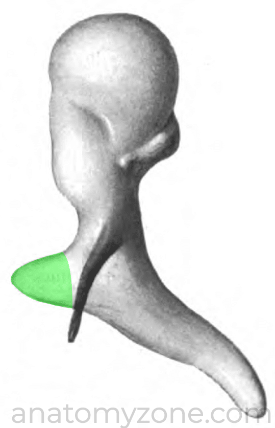
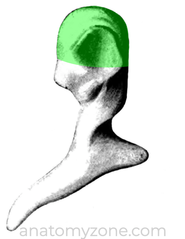
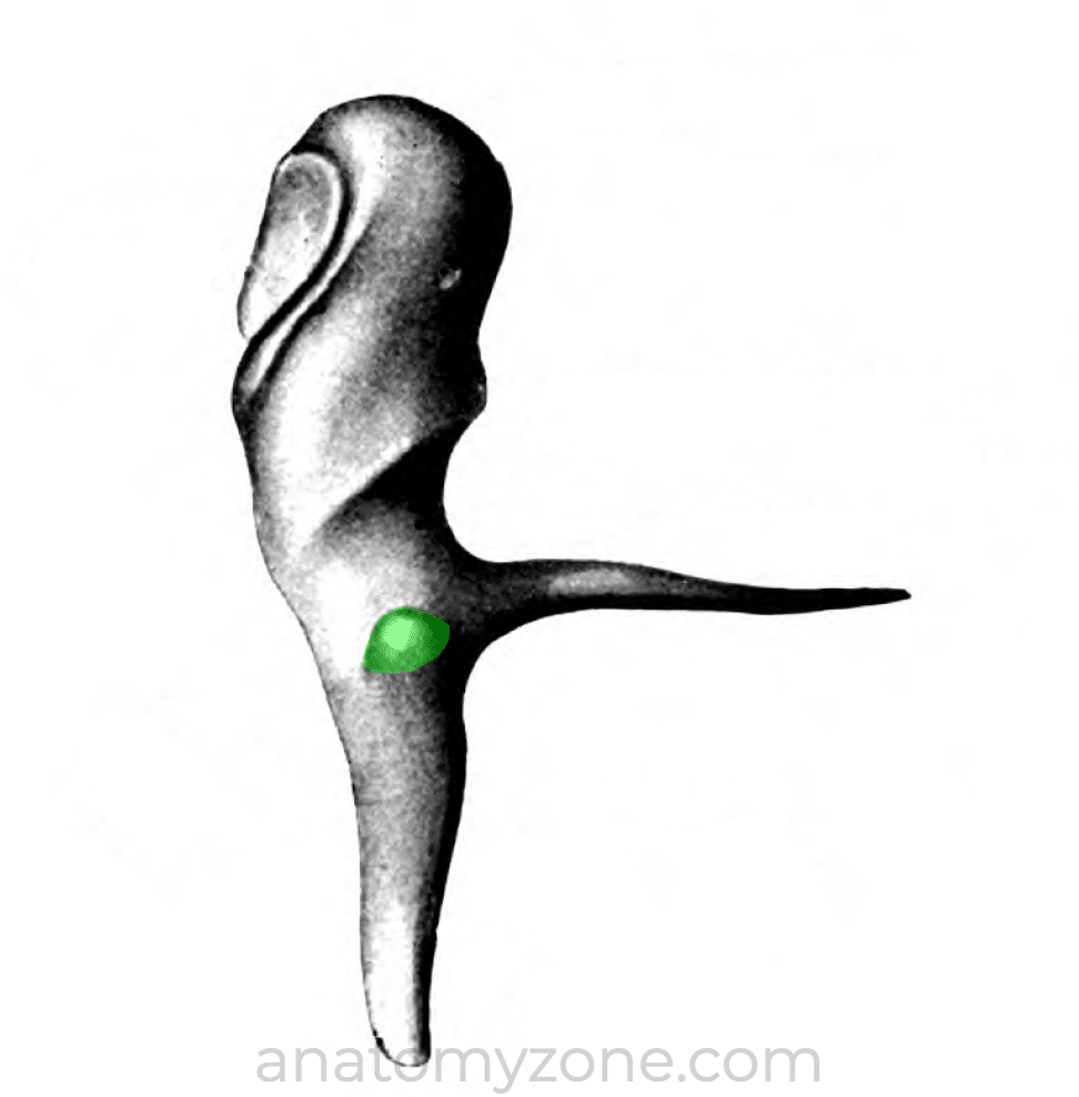
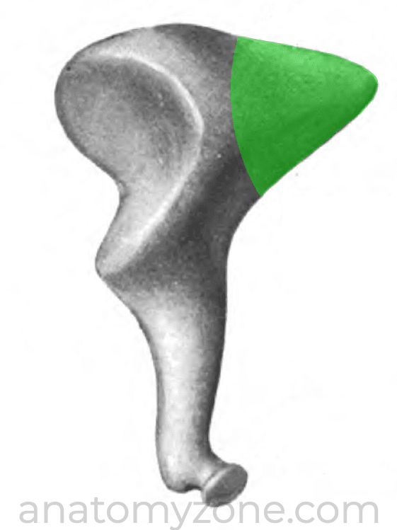
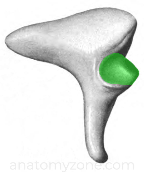
The head of the malleus is the rounded part which lies in the epitympanic recess superiorly and articulates with the incus via its posterior surface.
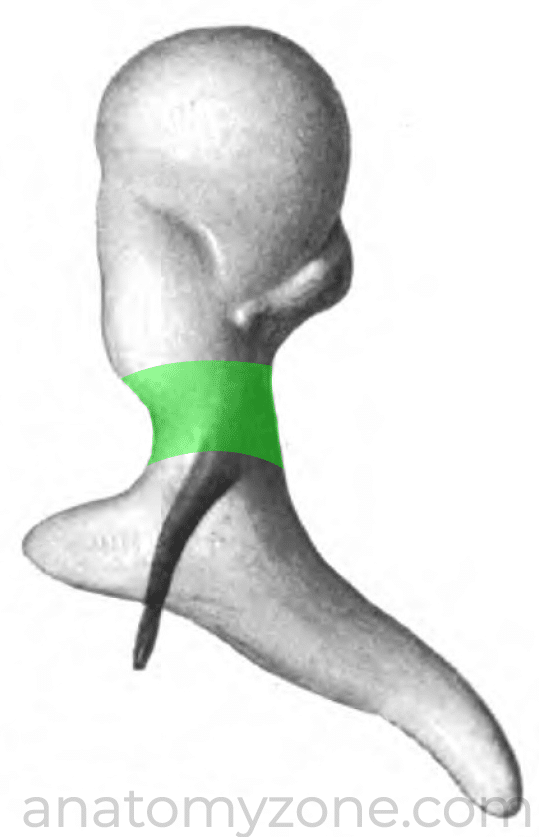
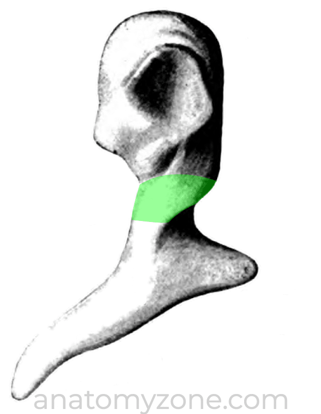
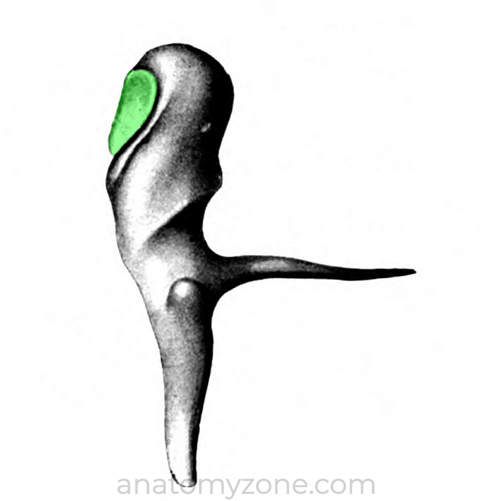
Just below the head, the malleus narrows to form the neck, which lies against the flaccid part of the tympanic membrane. The chorda tympani crosses the medial aspect of the malleolar neck.
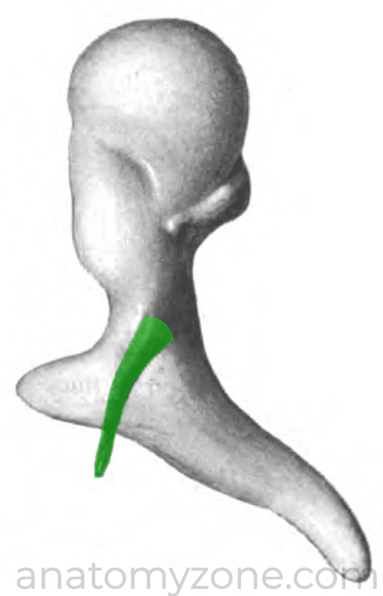
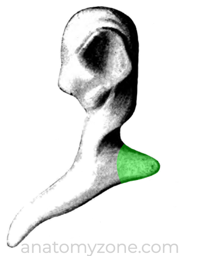
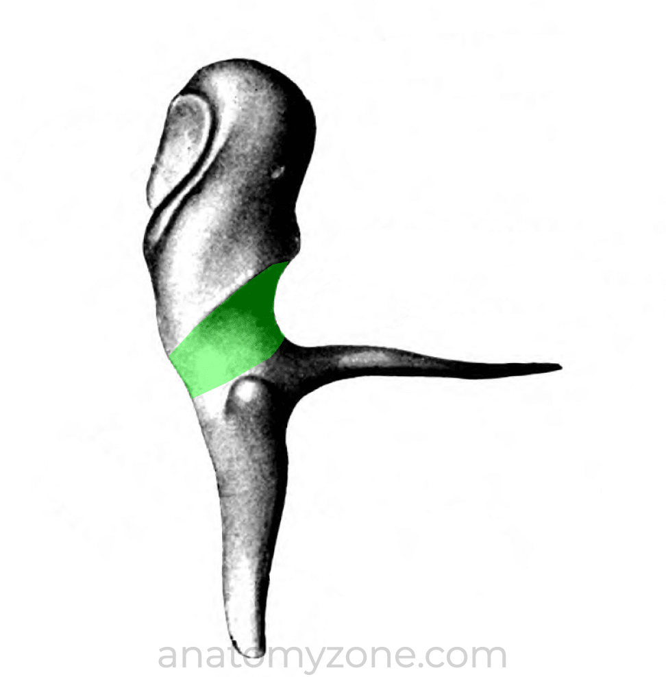
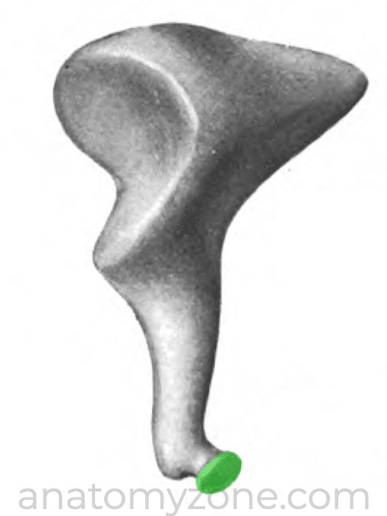
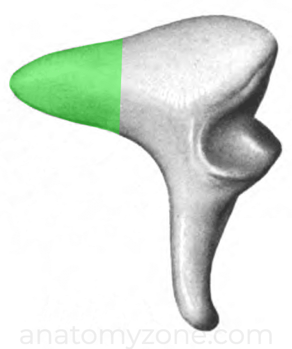
Below the neck of the malleus are the anterior and lateral processes of the malleus. The anterior process attaches to anterior wall of the tympanic cavity, and the lateral process attaches to the anterior and posterior malleolar folds of tympanic membrane.
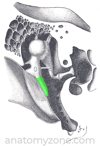
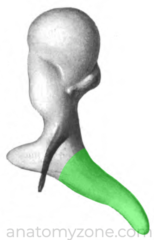
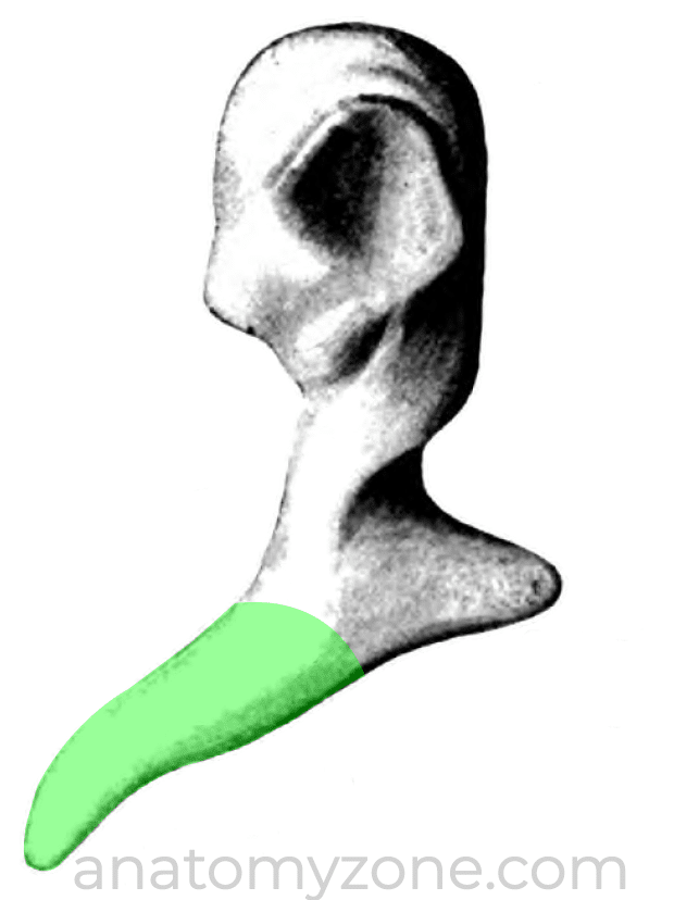
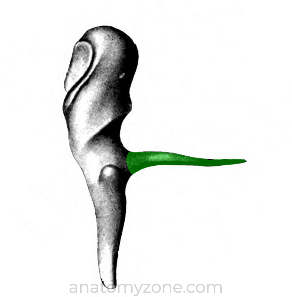
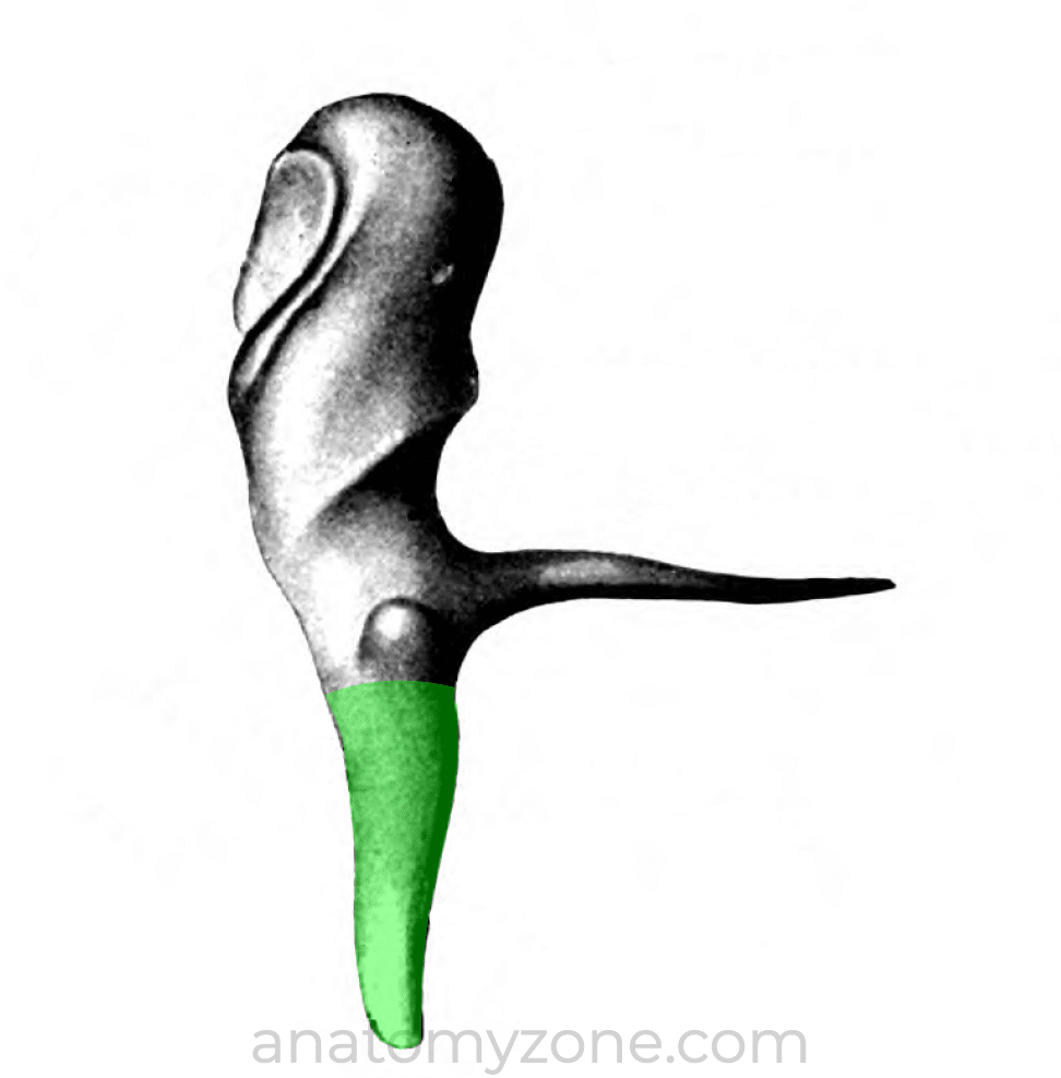
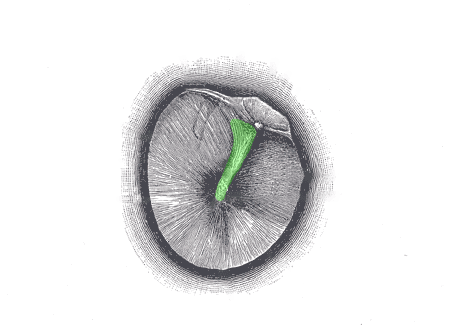
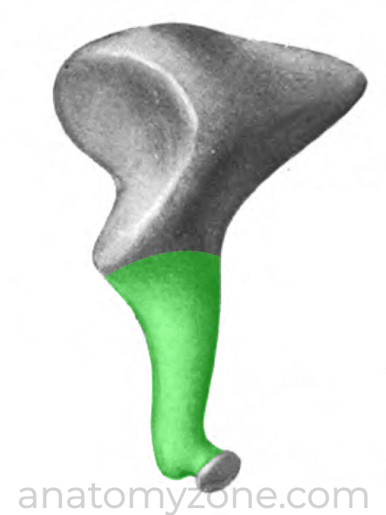
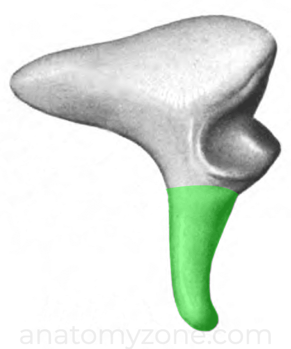
The inferior extension of the malleus below the anterior and lateral processes is known as the handle of malleus, or manubrium, which attaches to the tympanic membrane along the length of its lateral margin, with its tip at the umbo.
Incus
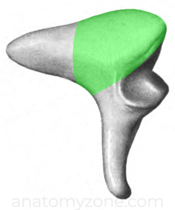
The incus is the second bone in ossicular chain, lying between the malleus and the stapes. It is comprised of a body and two limbs.
The body articulates with head of malleus at the incudomalleolar joint, and sits within the epitympanic recess.
The long limb extends inferiorly from the body, running in parallel to the handle of the malleus. Distally it curves medially to articulate with the stapes at the incudostapedial joint, via a small ovoid process known as the lenticular process.
The short limb extends posteriorly, attaching the incus to the posterosuperior wall of the middle ear.
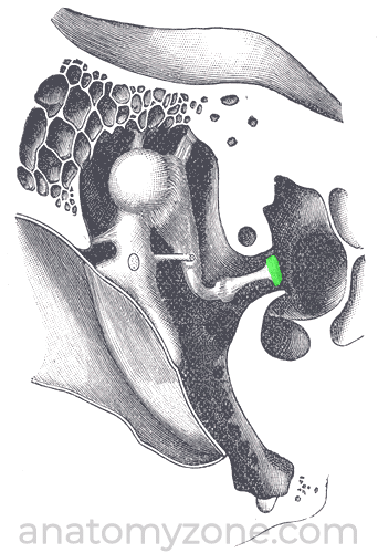
Stapes
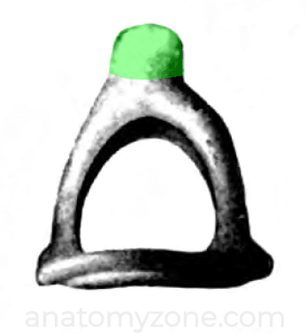
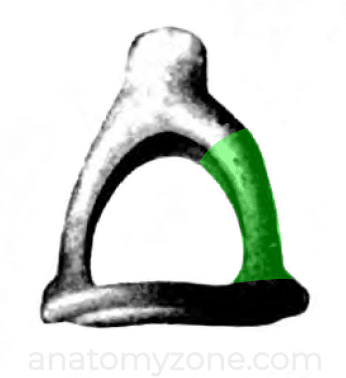
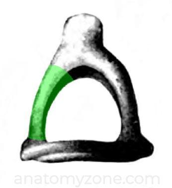
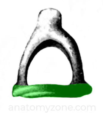
The stapes is the smallest of the ossicles, and the smallest bone in the human body and is comprised of a head, neck, two limbs and a footplate.
The head of the stapes is concave and articulates with the long limb of the incus via the lenticular process.
Just beneath the head is the neck of the stapes which is connected to the footplate or base of stapes by the anterior and posterior limbs.
The base, or stapes footplate attaches to the oval window on the medial wall of the middle ear.
Ossicular Joints
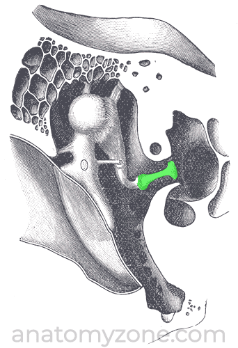
The articulations between the ossicles are synovial joints. Between the malleus and incus is a saddle-shaped synovial joint, and at the joint between the incus and stapedius is a ball and socket joint. As with other synovial joints elsewhere within the body, the articular surfaces are covered with cartilage and the joint is surrounded by a capsule lined by synovium.
Muscles of the Middle Ear
There are two muscles associated with the ossicular chain, the tensor tympani and the stapedius. These muscles contract in response to loud noises, reducing the amount that the ossicles vibrate and subsequently reducing transmission of sound to the inner ear structures.
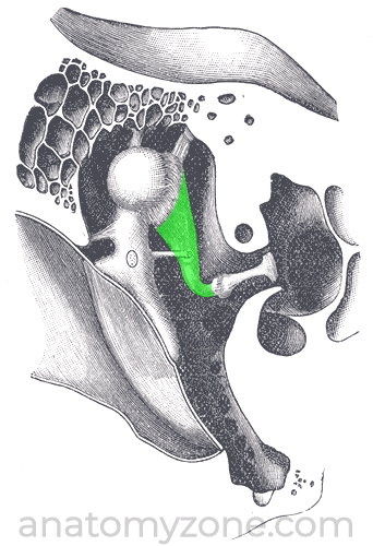
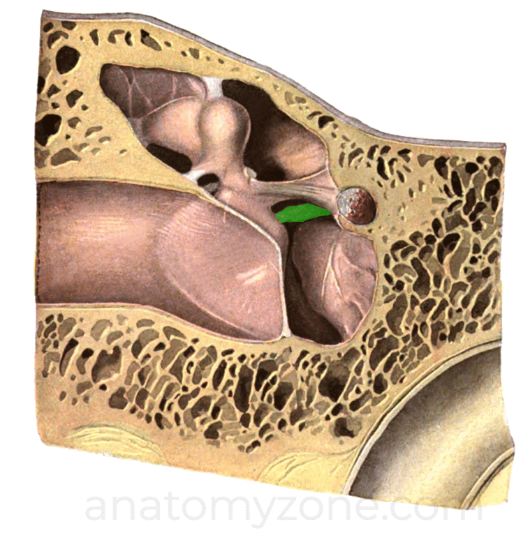
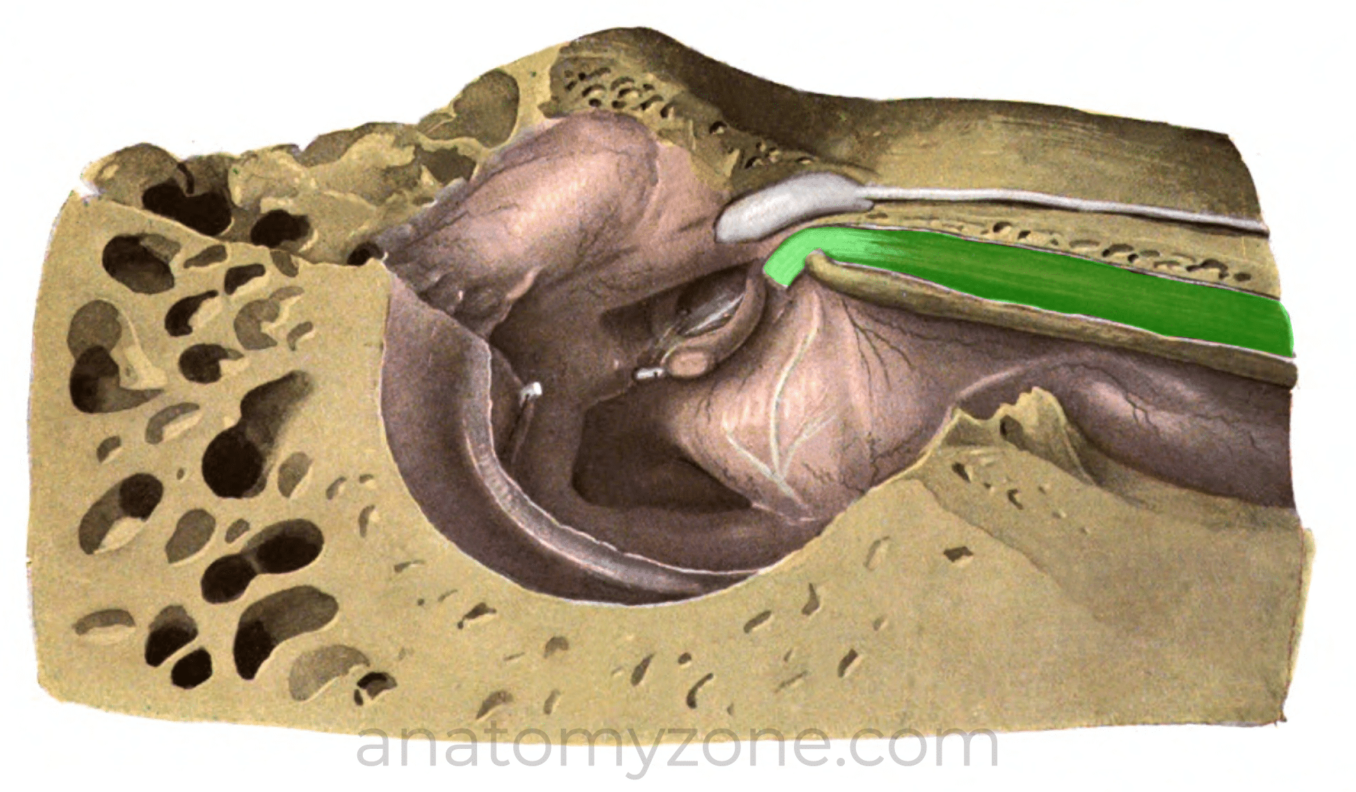
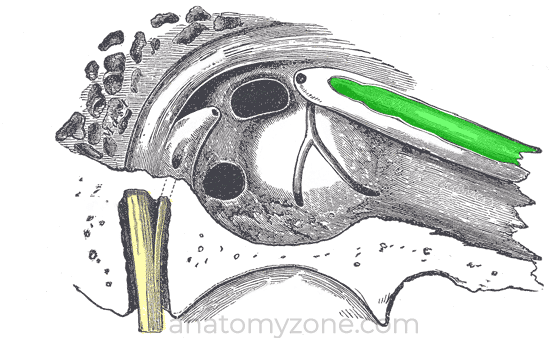
Tensor tympani
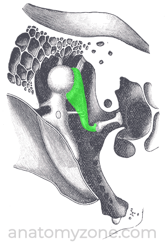
The tensor tympani lies within a bony canal parallel to the eustachian tube
Origin – cartilaginous part of eustachian tube, greater wing of sphenoid, and bony canal
Insertion – handle of malleus
Action – pulls handle of malleus medially to tense the tympanic membrane; reduces the force of vibrations.
Innervation – Branch of the mandibular nerve (V3)
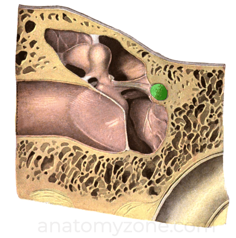
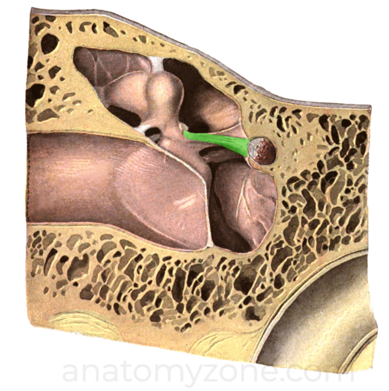
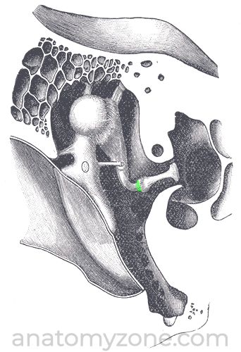
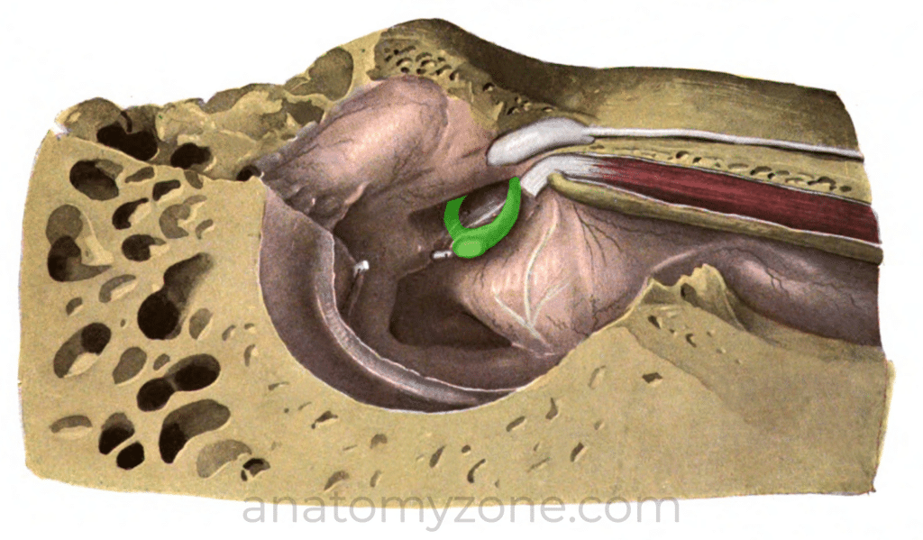
Stapedius
Origin – inside the pyramidal eminence (small protrusion on the posterior wall of the middle ear)
Insertion – Neck of stapes; tendon emerges from the apex of the pyramidal eminence.
Action – Pulls the stapes posteriorly, preventing excessive vibration in response to loud noise.
Innervation – Branch of the facial nerve (VII)

Ligaments of the Middle Ear
The ossicles are attached to the walls of the middle ear by various ligaments.
There are three ligaments for the malleus, and two for the incus and one for the stapes.
The ligaments attaching to the malleus include the anterior, posterior and lateral ligaments. The anterior ligament attaches from the neck of the malleus to the anterior wall of the middle ear. The lateral ligament attaches from the head of the malleus to the posterior part of tympanic notch. The superior ligament attaches from the head of the malleus to the roof of the epitympanic recess.
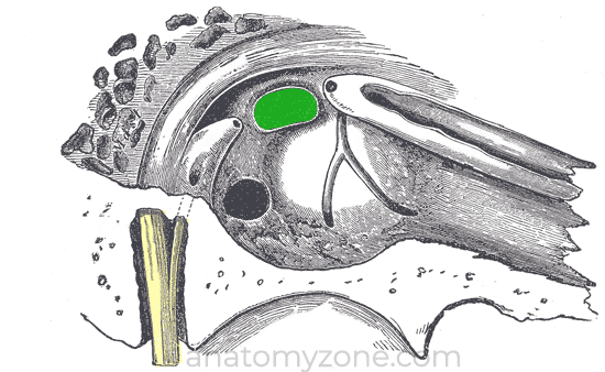
The posterior ligament of the incus attaches from the short limb to the fossa of incus (fossa incudis), which is a small depression in the posteroinferior aspect of the tympanic cavity. The superior ligament of incus may be considered as a small mucosal fold rather than a true ligament, and attaches from the body of the incus to the roof of the epitympanum.
The surface of the stapes footplate which attaches to the oval window is covered with hyaline cartilage and is attached to the oval window by a ring of fibres known as the annular ligament.