Introduction
The ear consists of three parts: external ear, middle ear and internal ear
The external ear consists of two parts:
- Auricle (pinna) – projects from the side of the head
- External auditory meatus (external acoustic meatus) – canal which leads to the middle ear
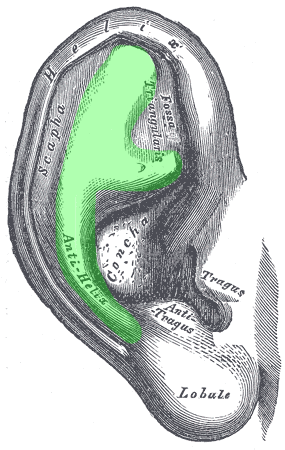
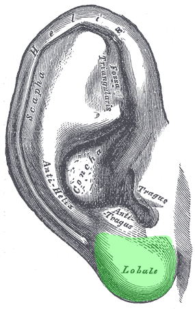
Auricle
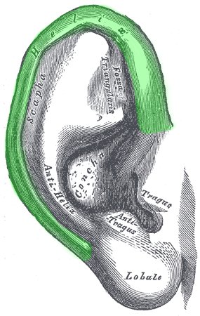
The auricle is a cartilaginous structure covered by skin which projects from the side of the head and is involved in capturing sound. It is compromised of the following parts:
- Helix – curved outer rim
- Antihelix – anterior to the helix. Crura form a ‘Y’ shape, with a superior crus and inferior crus.
- Lobule – fleshy non-cartilaginous inferior part of the auricle (“ear lobe”).
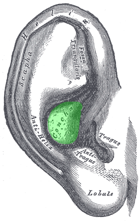
- Concha of auricle – centre of the auricle, external auditory meatus forms medially from here
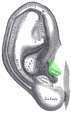
- Tragus – anterior to external auditory meatus
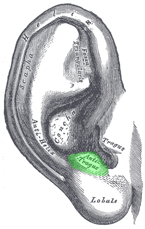
- Antitragus – opposite tragus, superior to lobule
Muscles
There are intrinsic and extrinsic muscles associated with the auricle.
The intrinsic muscles run through the cartilaginous framework of the auricle.
The extrinsic muscles consist of the anterior, superior and posterior auricular muscles.

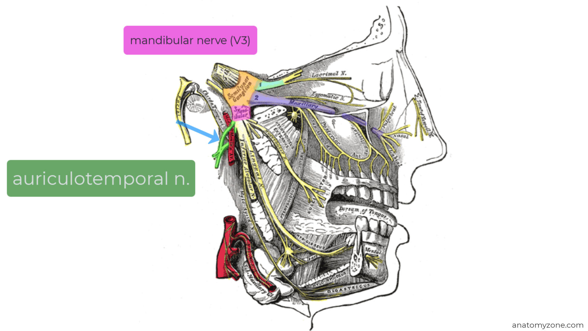
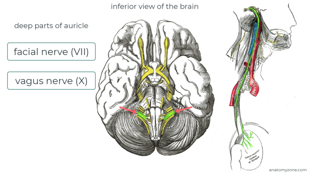
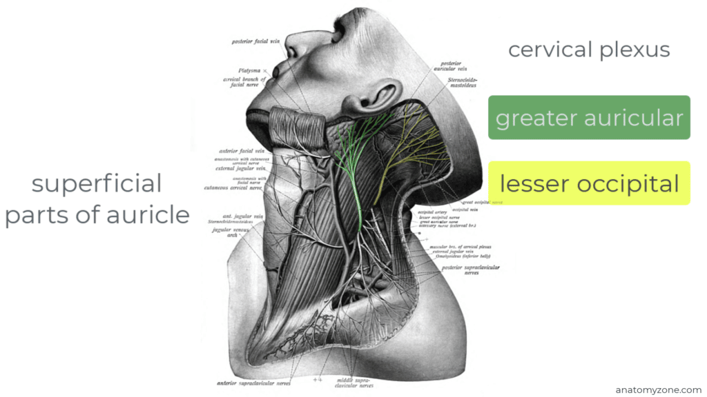
Innervation and Blood Supply to the Auricle
Several nerves provide sensory innervation to the auricle:
- Deep parts: branches of the facial nerve (Cn VII) and vagus nerve (X)
- Superficial/peripheral parts: greater auricular and lesser occipital nerves (from cervical plexus) and auriculotemporal branch of the mandibular nerve (Cn V3).
The arterial supply is from branches of the external carotid artery: posterior auricular, superficial temporal and occipital artery.
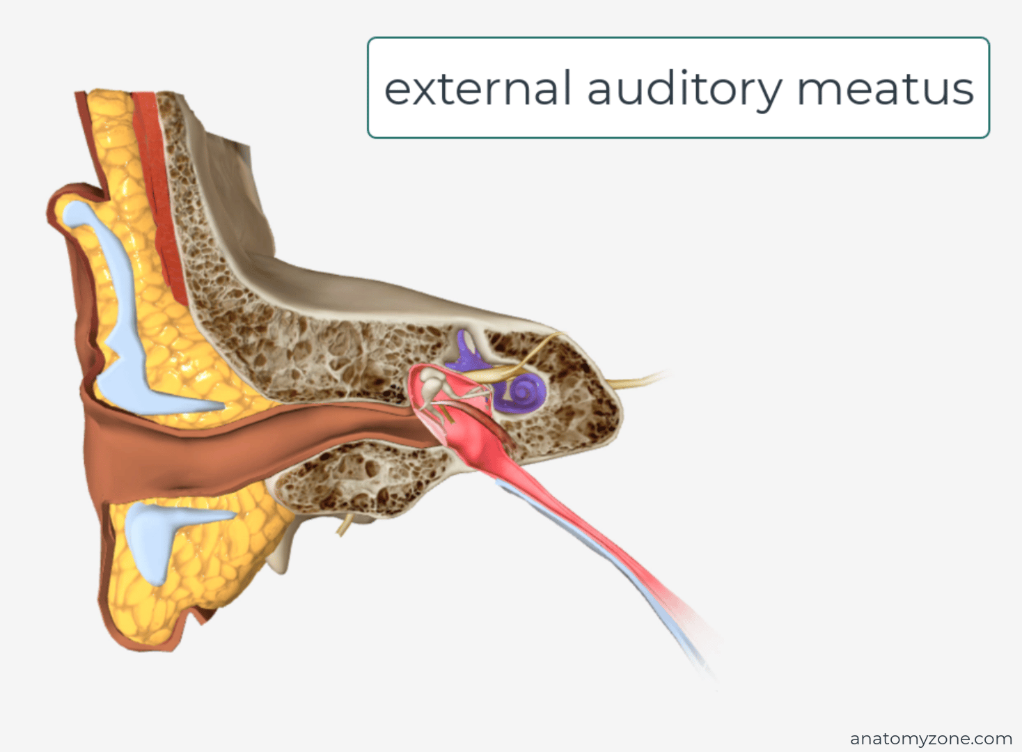
External Auditory Meatus
The external auditory meatus lies deep to the concha and extends medially to the tympanic membrane (ear drum).
- S-shaped course (not straight); auricle pulled up and back during otoscopic examination
- Cartilaginous lateral third (extension of auricular cartilage).
- Bony medial two thirds (temporal bone)
- Lined by skin along its course, contains small hair cells and specialised sweat gland which produce cerumen (ear wax).
- Sensory innervation: branches of auriculotemporal nerve, mandibular nerve and auricular branch of Vagus (X)
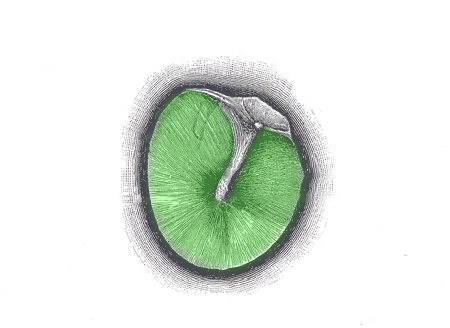
Tympanic membrane
The tympanic membrane is a thin membrane which separates the external ear from the middle ear compartment. It transmits sounds received via the external auditory meatus to the ossicles of the middle ear, via the oval window into the cochlear of the inner ear. It is attached to the temporal bone by a peripheral fibrocartilaginous ring and is angulated medially from superior to inferior and posterior to anterior.
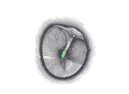
- Umbo – concavity where handle (manubrium) of malleus attaches
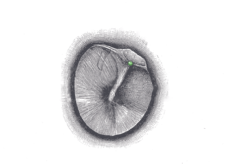
- Lateral process of malleus – superior to umbo and slightly anterior
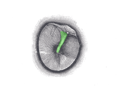
- Anterior and posterior malleolar folds – extends anteriorly/posteriorly from the lateral process of malleus
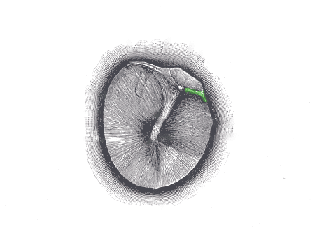
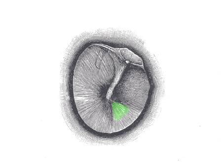
- Pars flaccida – small triangular area superior to malleolar folds; tympanic membrane thinner and loose
- Pars tensa – rest of the tympanic membrane; thicker and tighter
- Cone of light – seen anteroinferior to umbo on otoscopy
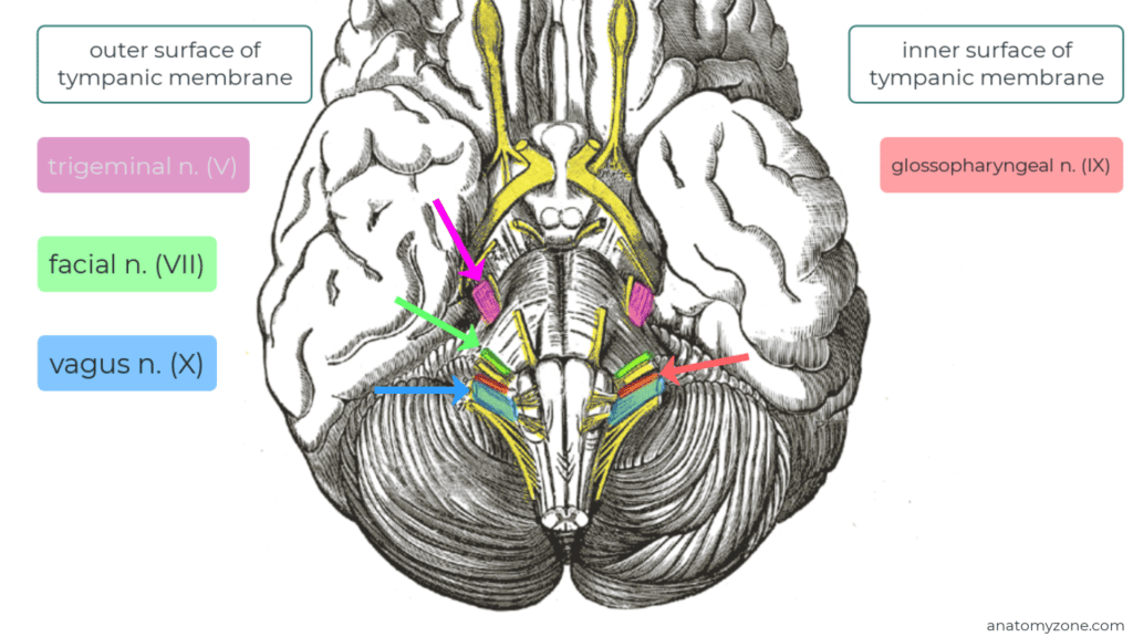
- Innervation: outer surface – trigeminal (V), facial (VII), vagus (X); inner surface – glossopharyngeal (IX)