Introduction
The middle ear is an air-filled space within the temporal bone which lies between the tympanic membrane laterally and the inner ear medially. It contains 3 ossicles which connect the tympanic membrane to the inner ear at the oval window. The ossicles transmit sound vibrations from the external auditory meatus into the inner ear apparatus. It is divided into two parts:
- Tympanic cavity (adjacent to the tympanic membrane)
- Epitympanic recess/attic (space superior to the tympanic cavity)
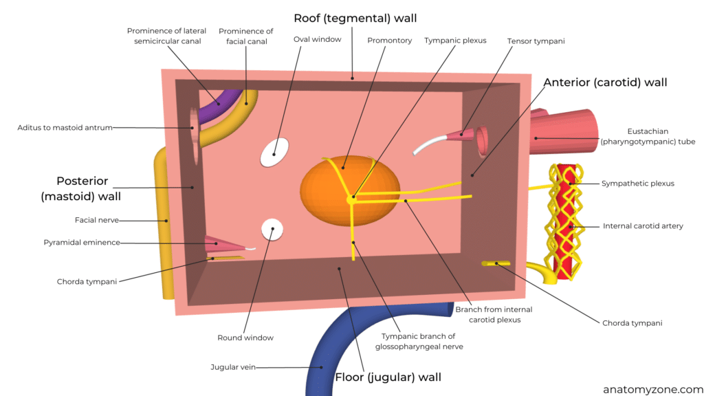
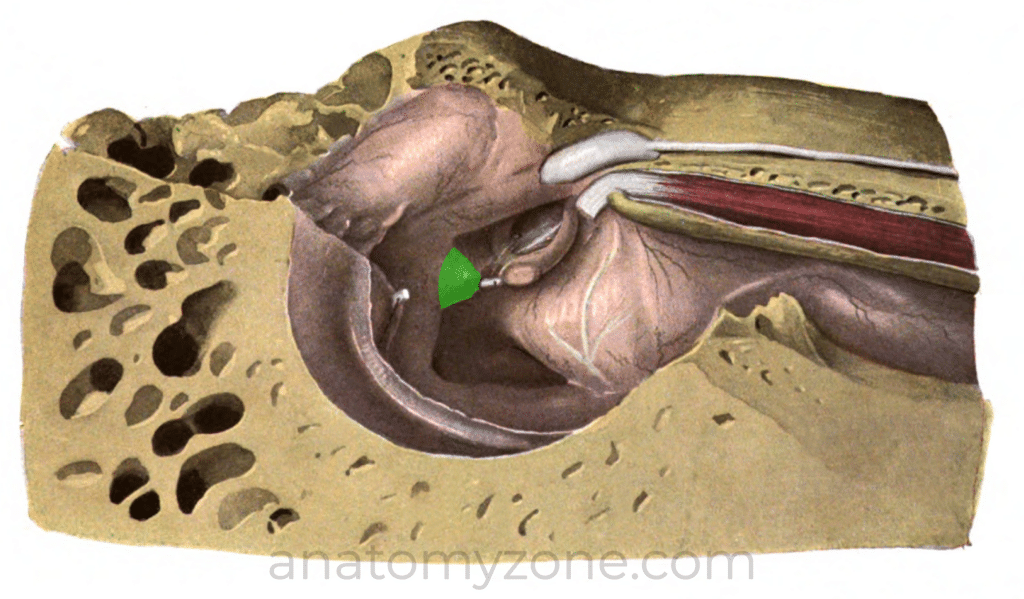
Medial (Labyrinthine) Wall
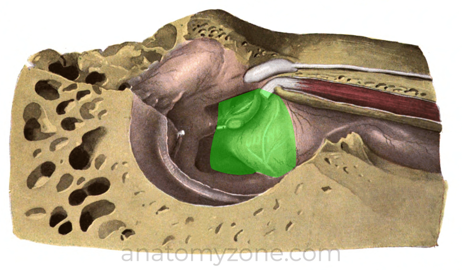
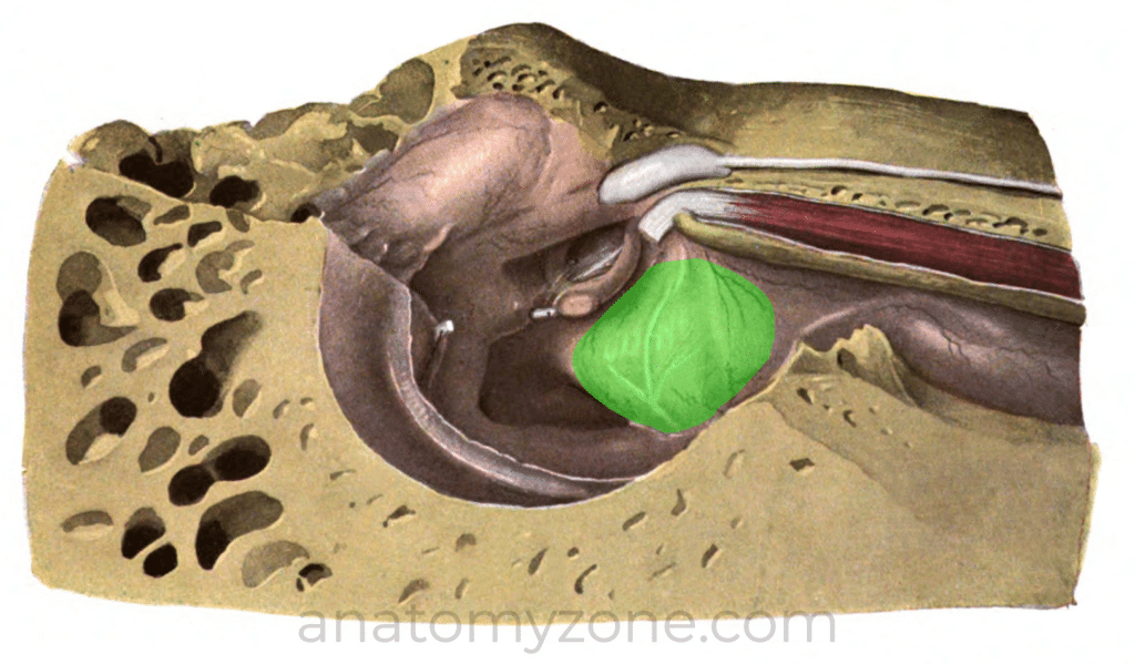
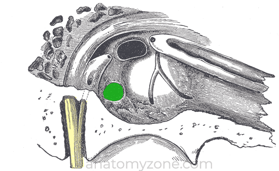
The medial (labyrinthine wall) is formed by the lateral aspect of the inner ear. The promontory is a convex protrusion formed by the bone overlying the basal turn of the cochlea.
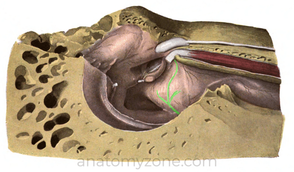
The promontory is covered by the tympanic plexus which receives branches from the glossopharyngeal nerve (IX) and branches of the internal carotid plexus.
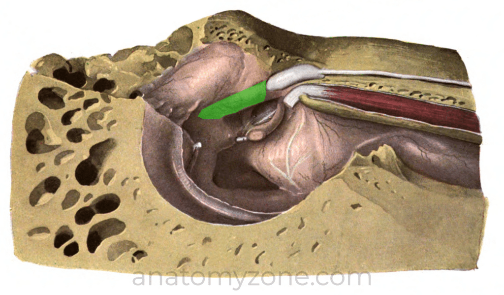
The medial wall contains 2 openings and two prominences.
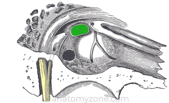
- 2 openings: Oval window – posterosuperior to the cochlear promontory and serves as the attachment for stapes footplate, which is the final point of attachment for ossicular chain. Round window – posteroinferior to cochlear promontory.
- Facial canal prominence: bony ridge formed by facial nerve canal
- Lateral semicircular canal prominence: sticks out laterally above the facial nerve canal
Lateral (Membranous) Wall
The lateral wall of the middle ear is formed mainly by the tympanic membrane. The superior aspect of the lateral wall is formed by the bony wall of the epitympanic recess
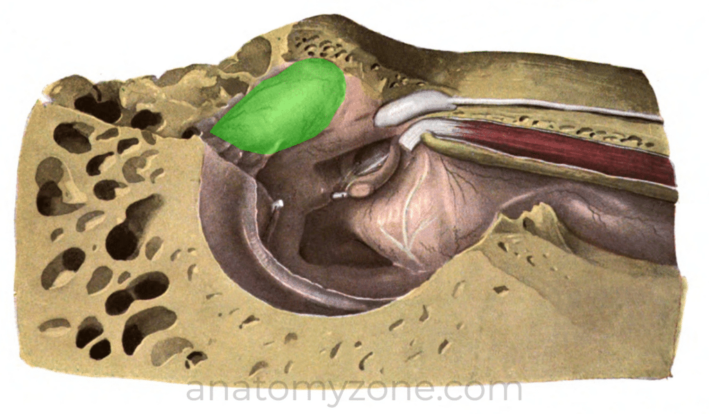
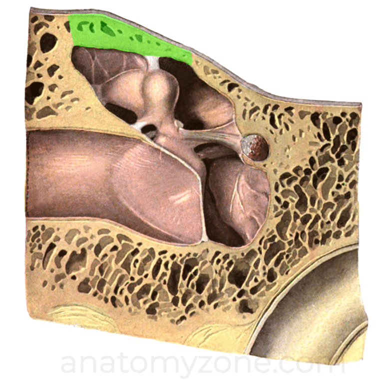
Roof (Tegmental Wall)
The roof separates the middle ear from the middle cranial fossa and is formed by a thin layer of bone known as the tegmen tympani.
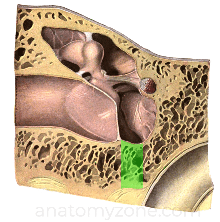
Floor (Jugular Wall)
The floor of the middle ear is formed by a thin layer of bone separating it from the internal jugular vein.
It contains a small aperture which transmits the tympanic branch from the glossopharyngeal nerve.
Posterior (Mastoid) Wall
Inferiorly within the posterior wall, there is a thin section of bone separating the middle ear cleft from mastoid air cells.
Superiorly within the posterior wall of the middle ear cavity is the aditus to mastoid antrum (continuous with the epitympanic recess). The pyramidal eminence is a small conical projection in front of the vertical segment of the facial nerve – it contains the stapedius muscle, and has a small opening through which the tendon passes into the middle ear. Also within the posterior wall, at the angle between the posterior and lateral walls, is the opening for the chorda tympani (the posterior canaliculus), which branches off from the mastoid segment of the facial nerve (VII). The chorda tympani then traverses the middle ear cavity and exits via the anterior canaliculus in the anterior wall.


Anterior (Carotid) Wall
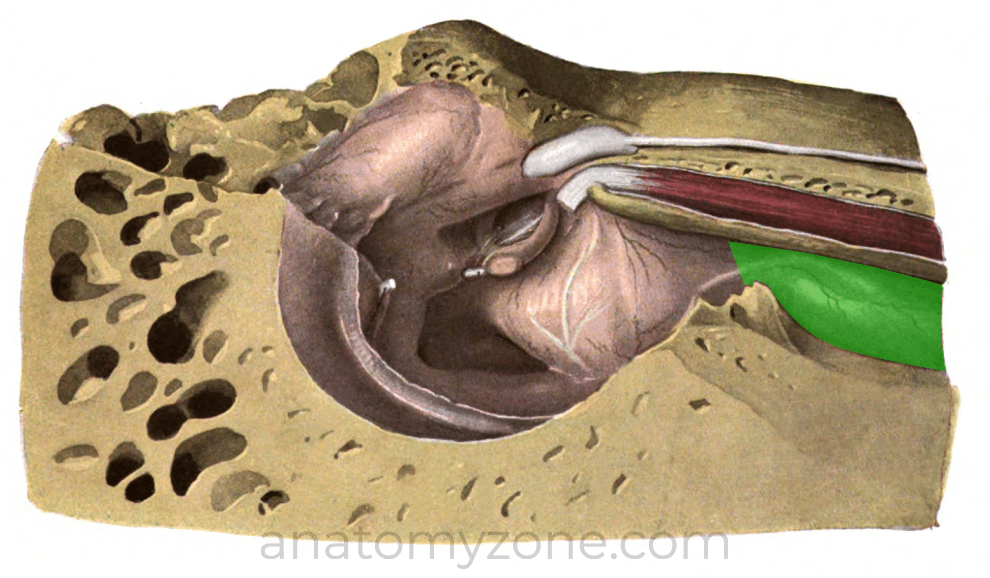
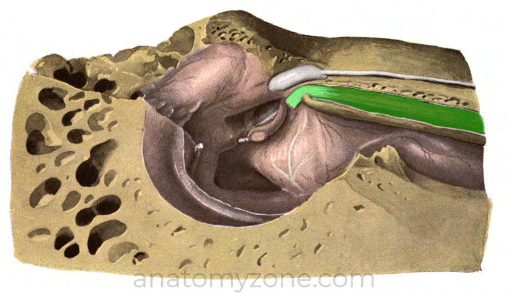
Inferiorly within the anterior wall is a thin layer of bone separating middle ear from the carotid canal. Superiorly it contains the opening of the eustachian tube (pharyngotympanic tube), and the opening for tensor tympani muscle (contained within a bony canal).
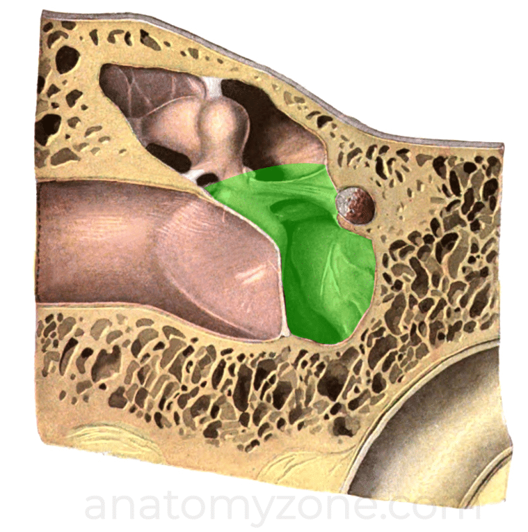
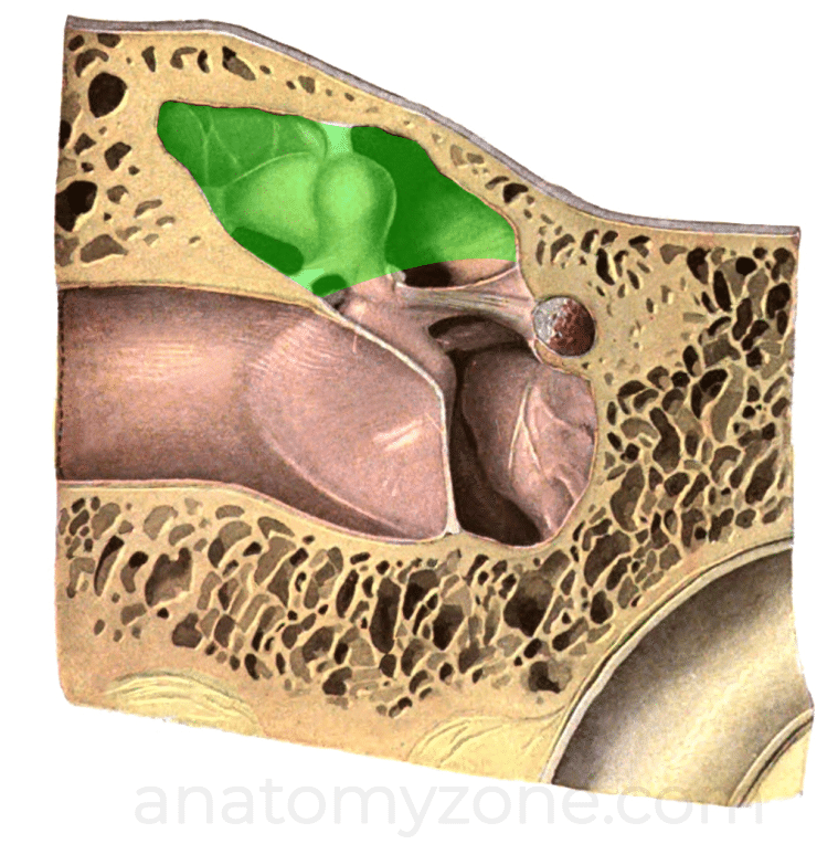
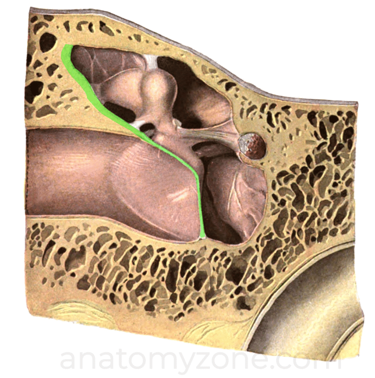
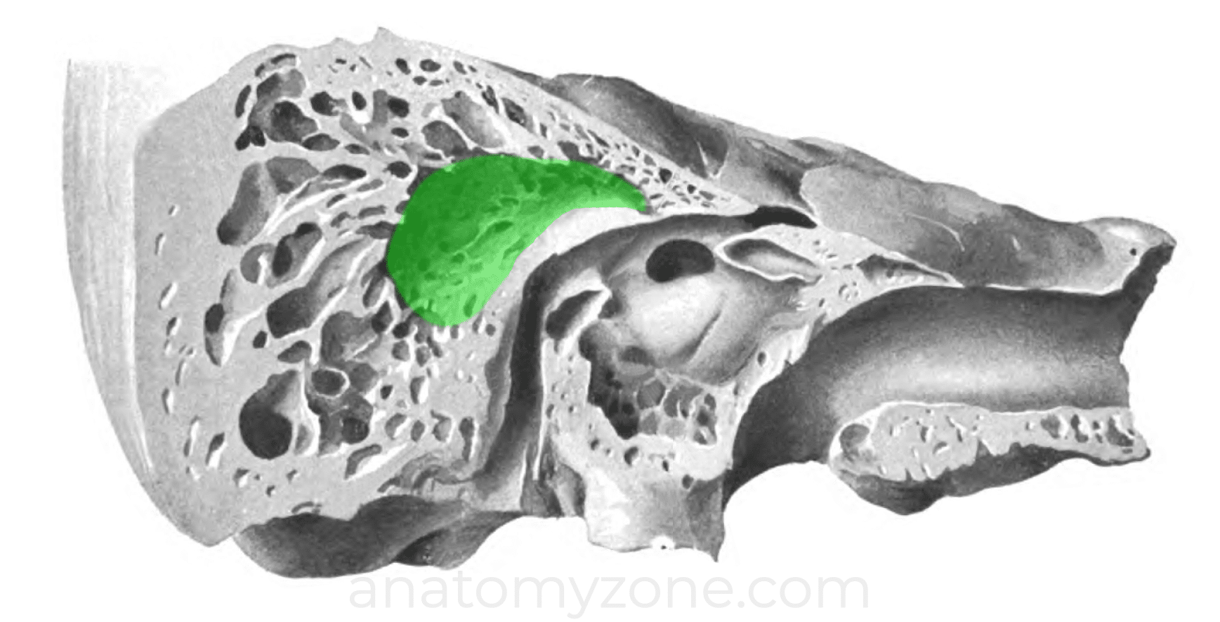
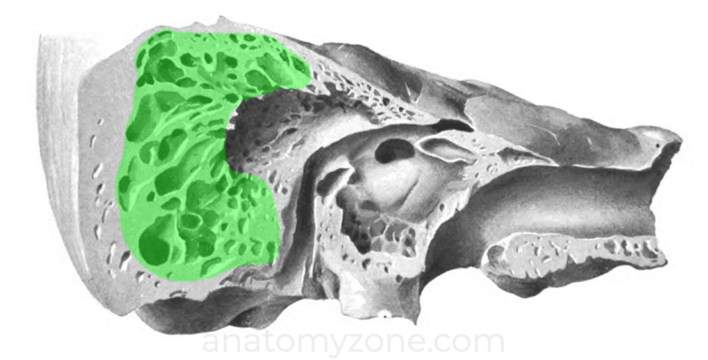
Mastoid Area
The mastoid area forms the posterior component of the temporal bone.
- Mastoid antrum: an irregularity cavity contained within the anterosuperior aspect of the mastoid area
- Aditus to mastoid antrum (aditus ad antrum): opening to the mastoid antrum, communicates anteriorly with the epitympanic recess
- Mastoid air cells: collections of air-filled spaces continuous with the mastoid antrum, extending into the mastoid process
- Tegmen mastoideum: the roof of the mastoid, which forms the posterior extension of the tegmen tympani. Separates the mastoid area from the middle cranial fossa.
- Sigmoid plate: thin lamina of bone separating mastoid air cells from the sigmoid sinus.
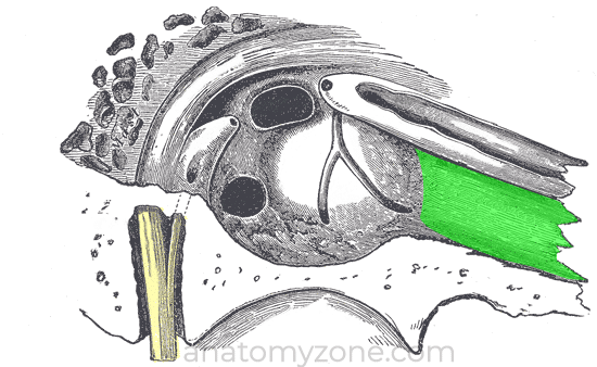
Eustachian Tube (Pharyngotympanic Tube)
The pharyngotympanic tube connects the middle ear with the nasopharynx, equalising pressure between the middle ear and external auditory meatus.
- Superior opening: anterior wall of the middle ear
- Inferior opening: nasopharynx
- Superior third – bony; inferior two thirds – cartilaginous