Internal Jugular Vein
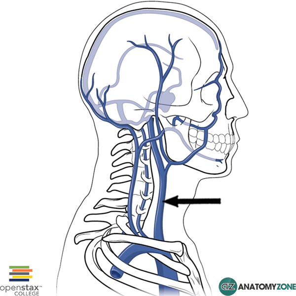
The structure indicated is the internal jugular vein.
The jugular veins are responsible for returning deoxygenated blood from the head to the heart via the superior vena cava. There are two jugular veins:
- External jugular vein
- Internal jugular vein
The external jugular vein drains into the subclavian vein. The internal jugular vein joins the subclavian vein to form the brachiocephalic vein. The brachiocephalic veins (innominate vein) on either side then join one another to form the superior vena cava, to bring blood into the right atrium of the heart. The external jugular vein runs superficial to the sternocleidomastoid muscle, the internal jugular vein on the other hand runs deep to this muscle inside the carotid sheath along with the common carotid artery and the vagus nerve.
Due to the direct pathway between the internal jugular vein and the right atrium of the heart (there is only a single pair of valves between the two), the internal jugular vein can be used as a “barometer” to measure the pressure in the venous system due to problems arising in the heart and lungs, resulting in problems with the right side of the heart. The jugular venous pressure (JVP) is the pulsation seen in the internal jugular vein, allowing one to estimate the pressure in the right atrium. It may be raised in conditions such as right ventricular failure.