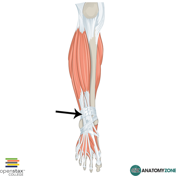
Superior Extensor Retinaculum
The structure indicated is the superior extensor retinaculum of the leg.
Retinacula (retinaculum singular) are bands of connective tissue which surround tendons and hold them in place. They function to stabilise tendons as the muscles to which they attach contract to cause movement. There are retinacula in the wrist, ankle and knee.
In the ankle, there is the flexor retinaculum, the peroneal retinacula and on the extensor surface, there is the superior and inferior extensor retinacula. The superior retinaculum is situated distally in the leg just above the ankle joint, attaching to the anterior aspects of the fibula and tibia.
The inferior retinaculum is Y-shaped. The base of the “Y” attaches laterally on the calcaneus. The upper arm of the “Y” attaches to the medial malleolus, whereas the lower arm of the “Y” attaches medially on the plantar aponeurosis.
The following tendons pass underneath the superior extensor retinaculum:
- Extensor digitorum longus tendon
- Extensor hallucis longus tendon
- Peroneus tertius tendon
- Tibialis anterior tendon
Learn more about the ankle joint in this tutorial.