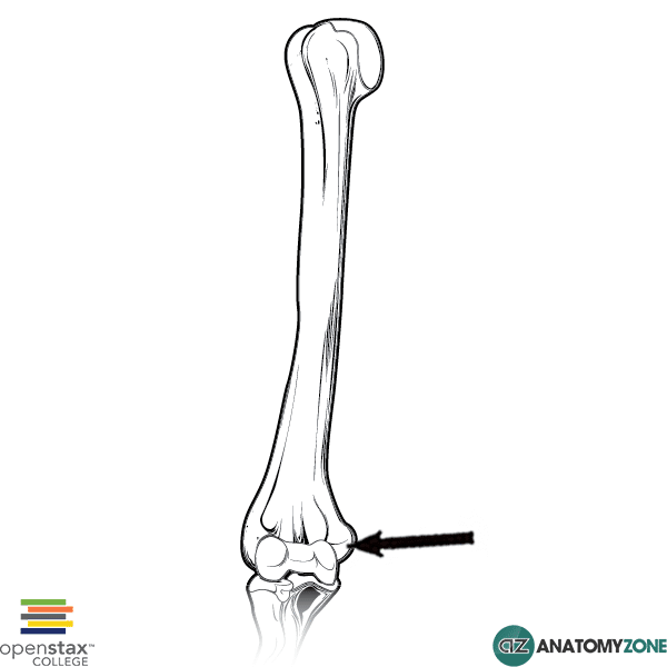
Medial Epicondyle of Humerus
The structure indicated is the medial epicondyle of the humerus.
The distal end of the humerus consists of several features:
- Condyle, consisting of the capitulum and trochlea
- Medial and lateral epicondyles
- Medial and lateral supracondylar ridges
- Radial fossa, coronoid fossa, olecranon fossa
A large central condyle which has two articular components – the capitulum which articulates with the radius, and the trochlea which articulates with the ulna. Either side of the humeral condyle, are two epicondyles, the medial and lateral epicondyles superior to which are the medial and lateral supracondylar ridges.
The medial and lateral epicondyles are easily palpable, and form the sites of origin for the forearm flexors of the anterior compartment and forearm extensors of the posterior compartment respectively. The medial epicondyle is a particularly important landmark, as the ulnar nerve passes around its posterior aspect to enter the forearm – it can easily be compressed or damaged at this location. The medial epicondyle is more prominent than the lateral epicondyle.
Learn more about the anatomy of the humerus in this anatomy tutorial.
