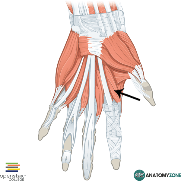
First Lumbrical
The structure indicated is the first lumbrical muscle of the hand.
The lumbricals belong to the intrinsic group of muscles which act on the hand. The intrinsic muscles include the following muscles/groups:
- Thenar group (act on thumb)
- Hypothenar group (act on little finger)
- Adductor pollicis
- Lumbricals
- Interossesus muscles
- Palmaris brevis
All the intrinsic muscles of the hand, except the thenar muscles and the lateral two lumbrical muscles are innervated by the deep branch of the ulnar nerve. The thenar muscles and the lateral two lumbrical muscles are innervated by the median nerve. A useful mnemonic for remembering this is MEATLOAF. “MEAT” refers to the Median nerve, and LOAF refers to the muscles which it innervates: Lateral two lumbricals, Opponens pollicis, Abductor pollicis brevis, Flexor pollicis brevis.
There are 4 lumbrical muscles which originate from the tendon of the flexor digitorum profundus muscle. The lateral lumbricals are unipennate, whereas the medial lumbricals are bipennate. The tendon of the lumbricals pass dorsally to insert onto the extensor expansions associated with the tendons of the extensor digitorum muscle. Because of the unique structure of the extensor expansions, the lumbrical muscles act to flex at the metacarpophalangeal joint, and extend at the interphalangeal joint.
Origin: Tendons of flexor digitorum profundus
Insertion: Extensor expansions of digits 2-5
Action: Flexion of metacarpophalangeal joints. Extension of interphalangeal joint
Innervation: Lateral two lumbricals: median nerve. Medial two lumbricals: deep branch of ulnar nerve.
Learn more about the anatomy of the hand muscles in this tutorial.