Carpal Tunnel
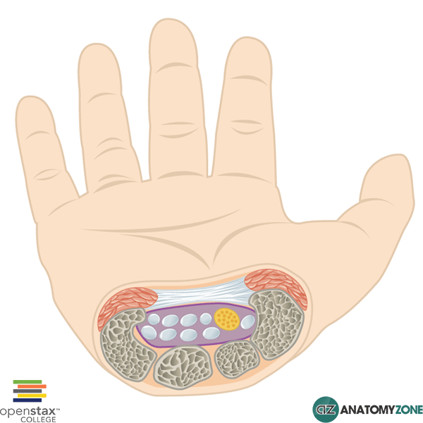
Represented in this diagram is the carpal tunnel.
The carpal tunnel is formed by an arch of carpal bones and the flexor retinaculum, found on the anterior aspect of the wrist. The flexor retinaculum attaches on the radial side to the scaphoid tubercle and the trapezium bone; on the ulnar side it attaches to the hook of the hamate and the pisiform. Its attachment over the arch of carpal bones converts the space into a tunnel which allows for the passage of several tendons and the median nerve.
The carpal tunnel contains nine long flexor tendons and the median nerve:
- Flexor digitorum profundus (four tendons)
- Flexor digitorum superficialis (four tendons)
- Flexor pollicis longus (one tendon)
The tendon of flexor carpi radialis passes through the flexor retinaculum in its own compartment – it does not technically pass through the carpal tunnel.
Carpal tunnel syndrome occurs due to pressure on the median nerve within the carpal tunnel. This typically causes sensations of pain/pins and needles in the distribution of the median nerve, as well as muscle weakness and atrophy of the thenar muscles.
Common causes of carpal tunnel syndrome include rheumatoid arthritis, pregnancy, hypothyroidism, acromegaly and trauma.