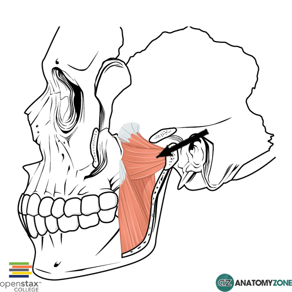
Lateral Pterygoid
The structure indicated is the lateral pterygoid muscle.
The lateral pterygoid muscle is one of four muscles of mastication:
- Masseter
- Temporalis
- Medial pterygoid
- Lateral pterygoid
The muscles of mastication act on the temporomandibular joint (TMJ), which is the joint of the jaw formed between the mandible and temporal bone. The TMJ is a synovial joint, which differs in structure to other synovial joints in that it contains a fibrocartilagenous articular disc that divides the joint into two parts. The two joint compartments result in different movements at the TMJ. The lower joint compartment allows rotational movement, whilst the upper joint compartment allows translational movement. There are four movements at the TMJ:
- Protrusion
- Retraction
- Elevation
- Depression
The lateral pterygoid muscle is located in the infratemporal fossa and consists of two heads:
- Upper head
- Lower head
Due to the primarily horizontal orientation of the fibres of the lateral pterygoid, its primary function is protrusion of the mandible.
Origin: Superior head: infratemporal crest of greater winger of sphenoid. Inferior head: lateral plate of the pterygoid process
Insertion: Conjoined tendon to neck of mandible
Action: bilateral contraction: protrusion of mandible. Unilateral contraction: side-side jaw movements
Innervation: mandibular branch of trigeminal nerve (cranial nerve V)
Learn more about the anatomy of the muscles of mastication in this anatomy tutorial.